| CAS NO: | 150347-59-4 |
| 包装 | 价格(元) |
| 10mg | 电议 |
| 25mg | 电议 |
| Storage | Store at -20°C |
| M.Wt | 557.46 |
| Cas No. | 150347-59-4 |
| Formula | C29H19NO11 |
| Solubility | ≥37.17 mg/mL in DMSO with ultrasonic; insoluble in EtOH; insoluble in H2O |
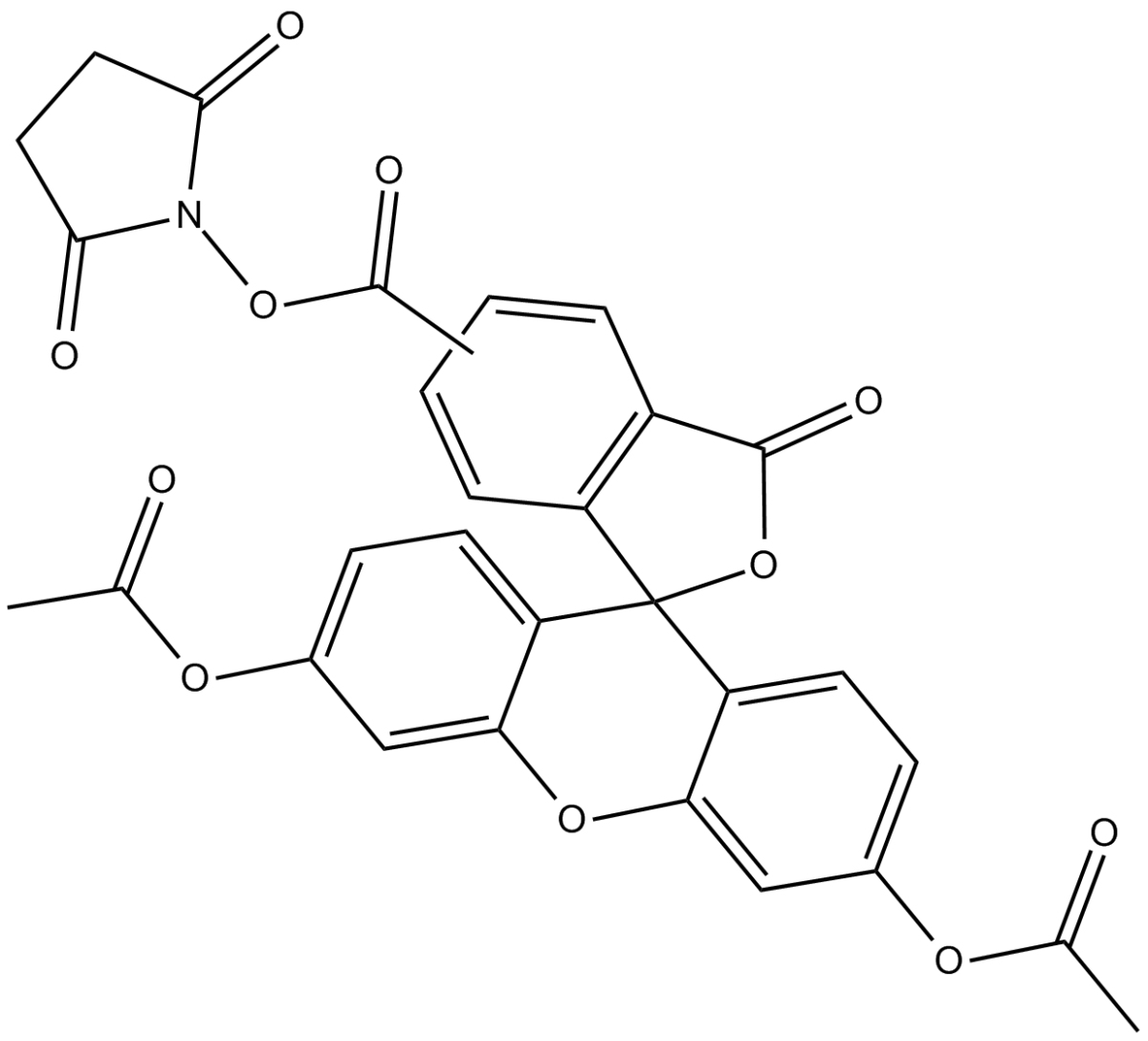
| Chemical Name | 3',6'-bis(acetyloxy)-3-oxo-2,5-dioxo-1-pyrrolidinyl ester-spiro[isobenzofuran-1(3H),9'-[9H]xanthene]-ar-carboxylic acid |
| Canonical SMILES | CC(OC1=CC2=C(C3(C4=C(C=C(OC(C)=O)C=C4)O2)C5=C(C(O3)=O)C=C(C(ON6C(CCC6=O)=O)=O)C=C5)C=C1)=O.CC(OC7=CC8=C(C9(C%10=C(C(O9)=O)C=CC(C(ON%11C(CCC%11=O)=O)=O)=C%10)C%12=C(C=C(OC(C)=O)C=C%12)O8)C=C7)=O |
| 运输条件 | 蓝冰运输或根据您的需求运输。 |
| 一般建议 | 为了使其更好的溶解,请用37℃加热试管并在超声波水浴中震动片刻。不同厂家不同批次产品溶解度各有差异,仅做参考。若实验所需浓度过大至产品溶解极限,请添加助溶剂助溶或自行调整浓度。溶液形式一般不宜长期储存,请尽快用完。 |
CFDA SE,全称为Carboxyfluorescein diacetate, succinimidyl ester,具有细胞膜通透性,一种广泛用于活细胞示踪或细胞增殖检测的荧光染料。CFDA SE本身不发光,进入活细胞后可以被细胞内的酯酶催化分解成CFSE,CFSE可发强烈的绿色荧光,后者不能穿透细胞膜,能完好的保留在细胞内。CFSE可以偶发性地并不可逆地和细胞内蛋白的Lysine残基或其它氨基发生结合反应,从而标记上这些蛋白。被CFDA SE标记的非分裂细胞的荧光非常稳定,稳定标记的时间可达数个月。
CFDA SE标记细胞的荧光非常均匀和稳定,荧光可平均分配至两个子代细胞中[1],这样每分裂一次子代细胞的荧光会减弱一半,通过流式细胞仪检测就可以检测出没有分裂的细胞,分裂一次的细胞(1/2的荧光强度),分离两次的细胞(1/4的荧光强度),分裂三次的细胞(1/8的荧光强度)以及类似的更多分裂次数的细胞。使用CFDA SE检测可以提供整个细胞群中有多少比例的细胞分裂了1次、2次或更多次数,可以检测分裂多达8次或更多次数的细胞增殖[2]。
目前CFDA SE标记细胞后通常用流式细胞仪进行细胞增殖检测。最常用于淋巴细胞的增殖检测,也可以用于成纤维细胞、NK细胞等其它细胞的增殖检测,甚至还可以用于细菌增殖的检测。CFDA SE标记细胞呈绿色荧光,Ex=494 nm,Em=521 nm。使用流式细胞仪检测时可以采用FL1 detection channel。CFDA SE标记的细胞也可以用荧光显微镜进行观察。CFDA SE标记的细胞可用于体外和体内增殖研究,且都不会使邻近细胞染色[3]。
References:
[1] Azarsiz E, Karaca N, Ergun B, et al. In vitro T lymphocyte proliferation by carboxyfluorescein diacetate succinimidyl ester method is helpful in diagnosing and managing primary immunodeficiencies. J Clin Lab Anal. 2018;32(1):e22216.
[2] Lyons AB, Blake SJ, Doherty KV. Flow cytometric analysis of cell division by dilution of CFSE and related dyes. Curr Protoc Cytom. 2013;Chapter 9:Unit9.11.
[3] Li X, Dancausse H, Grijalva I, et al. Labeling Schwann cells with CFSE-an in vitro and in vivo study. J Neurosci Methods. 2003 May 30;125(1-2):83-91.
| Cell experiment:[1] | |
Cell lines | Human erythroleukaemic cell line K562, mouse lymphoma cell line YAC-1, human mammary cancer cell line MCF-7 and human melanoma cell line A375 |
Reaction Conditions | 1, 1.5, 2, 2.5, 5 and 10 μM CFDA-SE |
Applications | CFDA-SE at 2.5 μM stained more than 95% of the cells on all cell lines tested, and dose-dependently increased fluorescence intensity of stained cells. The optimal concentration for K562 and YAC-1 was found to be 2.5 μM, while the optimal concentrations for A375 and MCF-7 were found to be 5 μM and 10 μM, respectively. Within the 6 h experiment period, no cytotoxicity related to CFDA-SE was observed. |
| Animal experiment:[4] | |
Animal models | C57BL/6 mice, aged 5 ~ 8 weeks |
Dosage form | 10 μM Injected into thymic lobe |
Applications | CFDA-SE, at 80 times the concentration used forin vitrolabeling, was nontoxic and labeled randomly approximately 15% of thymocytes 24 h after injection. The turnover rate of labeled thymic emigrants in the lymph nodes was in the order of 21 days. Thus, CFDA-SE may serve as a powerful tool in relatively long-term migration studies. |
Note | The technical data provided above is for reference only. |
References: 1. Wang XQ, Duan XM, Liu LH, et al. Carboxyfluorescein diacetate succinimidyl ester fluorescent dye for cell labeling. Acta Biochimica et Biophysica Sinica (Shanghai), 2005, 37(6): 379-385. 2. Weston SA, Parish CR. New fluorescent dyes for lymphocyte migration studies. Analysis by flow cytometry and fluorescence microscopy. Journal of Immunological Methods, 1990, 133(1): 87-97. 3. Parish CR, Glidden MH, Quah BJ, et al. Use of the intracellular fluorescent dye CFSE to monitor lymphocyte migration and proliferation. Current Protocols in Immunology, 2009, Chapter 4: Unit4.9. 4. Graziano M, St-Pierre Y, Beauchemin C, et al. The fate of thymocytes labeled in vivo with CFSE. Experimental Cell Research, 1998, 240(1): 75-85. | |
 m.cnreagent.com