| CAS NO: | 65673-63-4 |
| 规格: | ≥98% |
| 包装 | 价格(元) |
| 100mg | 电议 |
| 250mg | 电议 |
| 500mg | 电议 |
| 1g | 电议 |
| Molecular Weight (MW) | 409.23 |
|---|---|
| Formula | C17H17BrN2O5 |
| CAS No. | 65673-63-4 |
| Storage | -20℃ for 3 years in powder form |
| -80℃ for 2 years in solvent | |
| Solubility (In vitro) | DMSO: 82 mg/mL (200.4 mM) |
| Water:<1 mg/mL | |
| Ethanol: 82 mg/mL (200.4 mM) | |
| Solubility (In vivo) | 1% DMSO+30% polyethylene glycol+1% Tween 80: 30 mg/mL |
| Synonyms | HA-141, HA 141, HA141 |
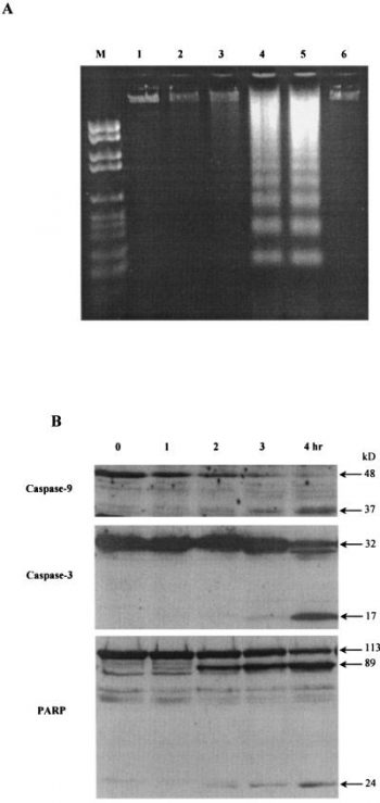
| In Vitro | Kinase Assay: The binding affinity of organic compounds to Bcl-2 protein in vitro is determined by a competitive binding assay based on fluorescence polarization. For this assay, 5-carboxyfluorecein is coupled to the N terminus of a peptide, GQVGRQLAIIGDDINR, derived from the BH3 domain of Bak (Flu-BakBH3), which has been shown to bind to the surface pocket of the Bcl-xL protein with high-affinity. According to our molecular modeling studies and binding measurement using fluorescence polarization, the Flu-BakBH3 peptide binds the surface pocket of Bcl-2 with a similar affinity. Bcl-2 used in this assay is a recombinant GST-fused soluble protein. Flu-BakBH3 and Bcl-2 protein are mixed in the presence or absence of organic compounds under standard buffer conditions and are incubated for 30 min. The binding of Flu-BakBH3 to Bcl-2 protein is measured on a LS-50 luminescence spectrometer equipped with polarizers using a dual path length quartz cell (500 μl). The fluorophore is excited with vertical polarized light at 480 nm (excitation slit width 15 nm), and the polarization value of the emitted light is observed through vertical and horizontal polarizers at 530 nm (emission slit width 15 nm). The binding affinity of each compound for Bcl-2 protein is assessed by determining the ability of different concentrations of the compound to inhibit Flu-BakBH3 binding to Bcl-2. Cell Assay: The cytotoxic effects of HA14-1 against different FL cell lines are determined by the MTT assay. Briefly, the cells (5000/well) are incubated in triplicate in 96-well plate in the presence or absence of HA14-1 for 20 h at 37 °C. Thereafter, the MTT solution is added to each well. After 4 h incubation at 37 °C, the optical density (OD) is measured by means of 96-well plate reader, with the extraction buffer as a blank. The following formula is used: percentage cell viability = (OD of the experiment samples/OD of the control) × 100. Sigmoidal dose-response curves are fitted to the mean cell viability plotted against log HA14-1 dose and lethal concentration 50% (LC50) values are calculated from the resulting curves using Prism software. HA14-1 is a small molecule and nonpeptidic ligand of a Bcl-2 surface pocket with IC50 of approximately 9 μM. HA14-1 induces the apoptosis of HL-60 cells in a dose-dependent manner via caspase activation. HA14-1 shows cytotoxic effects on HF1A3, HF4.9 and HF28RA follicular lymphoma B cell lines with LC50 of 4.5 μM, 12.6 μM and 8.1 μM respectively. HA14-1 induces apoptosis of HF1A3, HF4.9 and HF28RA cells via caspase and ROS. |
|---|---|
| In Vivo | HA14-1(400 nM) treatment did not have any significant effect on the growth of glioblastoma tumors in immunodeficient mice. But HA14-1 (400 nM) increases the effect of the DNA-damaging agent etoposide (2.5 mg/Kg) on glioblastoma growth in vivo. |
| Animal model | Female Swiss nude mice bearing BeGBM xenografts. |
| Formulation & Dosage | Formulated in Free RPMI 1640-50% DMSO.; 400 nM; Inject at the site of cell injection |
| References | Proc Natl Acad Sci U S A. 2000 Jun 20;97(13):7124-9.; Leuk Res. 2006 Mar;30(3):322-31. |
 Apoptosis induced by HA14-1 in HL-60 cells as indicated by DNA ladders. Proc Natl Acad Sci U S A. 2000 Jun 20; 97(13): 7124–7129. |  Effect of HA14-1 on Apaf-1–/– and Apaf-1+/+ cells. Apaf-1-deficient mouse embryonic fibroblast (Apaf-1–/–) and NIH 3T3 (Apaf-1+/+) cells were treated with 50 μM HA14-1 for 24 h. Percent cell death was determined by using Trypan blue staining. To examine the role of caspases in the death of Apaf-1+/+ cells induced by HA14-1, cells were preincubated with zVAD-fmk for 2 h before the treatment of the compound. |
