| 包装 | 价格(元) |
| 10mM (in 1mL DMSO) | 电议 |
| 50mg | 电议 |
Cell experiment: | MTT is used as an indicator of cell viability. Primary human umbilical vein endothelial cells (HUVECs) are grown in 96-well plates at a density of 5×10³ cells/well. After 24 h, the cells are washed with fresh medium and treated with MTU (0-20 μM). After a 48 h incubation period, the cells are washed, and 100 μL of MTT (1 mg/mL) is added, followed by incubation for 4 h. Finally, DMSO (150 μL) is added to solubilize the formazan salt formed, and the amount of formazan salt is determined by measuring the OD at 540 nm using a microplate reader [1]. |
Animal experiment: | Mice[1]Male C57BL/6 mice (6-7 weeks old; average weight, 27 g) are used in this study. Mice are administered LPS (0.3 mg/mouse or 15 mg/kg, intravenously). After 4 h, the mice are intravenously treated with MTU (142 or 284 μg/kg, for 6 h) and injected with 1% Evans blue dye solution in normal saline. Six hours later, the mice are sacrificed and peritoneal exudates are collected by washing cavities with 5 mL of normal saline and by centrifuging at 200× g for 10 min. The absorbance of the supernatant is read at 650 nm. Vascular permeabilities are expressed as μg of dye/mouse that leaked into the peritoneal cavity and are determined using a standard curve. |
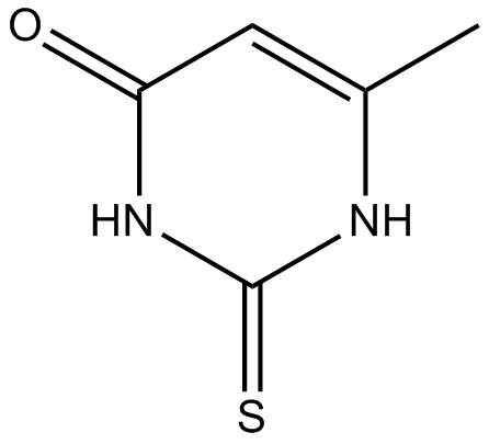
| 产品描述 | Methylthiouracil is an antithyroid agent. Methylthiouracil suppresses the production TNF-α and IL-6, and the activation of NF-κB and ERK1/2. HUVECs are treated with various concentrations of MTU (0-20 μM) for 6 h after the addition of LPS (100 ng/mL) for 4 h. MTU inhibits LPS-mediated hyperpermeability in endothelial cells, with the optimal effect occurring at a concentration above 5 μM. The effects of MTU are examined on HUVEC actin cytoskeletal arrangement by immunofluorescence staining of HUVEC monolayers with F-actin labeled fluorescein phalloidin. Control HUVECs exhibit a random distribution of F-actin throughout the cells, with some localization of actin filament bundles at the cell boundaries. Barrier disruption by LPS (100 ng/mL) is manifested by the formation of paracellular gaps in HUVECs. In addition, post-treatment with MTU (10 or 20 μM) results in inhibited formation of LPS-induced paracellular gaps with the formation of dense F-actin rings. To test the cytotoxicity of MTU, cellular viability assays are performed in HUVECs treated with MTU for 24 h. At concentrations up to 20 μM, MTU does not affect cell viability[1]. MTU treatment results in marked inhibition of the peritoneal leakage of dye induced by LPS. The average circulating blood volume for mice is 72 mL/kg. Because the average mouse weight in this study is 27 g, and the average blood volume is 2 mL, the injected MTU (142 or 284 μg/kg) results in a maximum concentration of 10 or 20 μM in the peripheral blood[1]. References: |
 m.cnreagent.com