| CAS NO: | 152044-53-6 |
| 规格: | ≥98% |
| 包装 | 价格(元) |
| 5mg | 电议 |
| 10mg | 电议 |
| 25mg | 电议 |
| 50mg | 电议 |
| 100mg | 电议 |
| 250mg | 电议 |
| 500mg | 电议 |
| Molecular Weight (MW) | 493.66 |
|---|---|
| Formula | C26H39NO6S |
| CAS No. | 152044-53-6 |
| Storage | -20℃ for 3 years in powder form |
| -80℃ for 2 years in solvent | |
| Solubility (In vitro) | DMSO: 99 mg/mL (200.5 mM) |
| Water: <1 mg/mL | |
| Ethanol: 99 mg/mL (200.5 mM) | |
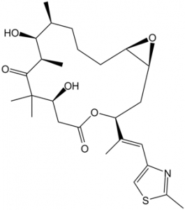
| SMILES | O=C(C(C)(C)[C@@H](O)C1)[C@H](C)[C@@H](O)[C@@H](C)CCC[C@@]2([H])O[C@@]2([H])C[C@@H](/C(C)=C/C3=CSC(C)=N3)OC1=O |
| Synonyms | Epothilone-A; EpoA; (1R,5S,6S,7R,10S,14S,16S)-6,10-Dihydroxy-5,7,9,9-tetramethyl-14-[(E)-1-(2-methyl-1,3-thiazol-4-yl)prop-1-en-2-yl]-13,17-dioxabicyclo[14.1.0]heptadecane-8,12-dione |
| In Vitro | In vitro activity: Epothilone A, discovered from the myxobacterium Sorangium cellulosum, is a Taxol-like microtubule-stabilizing agent that induces tubulin polymerization, leading to cell cycle arrest at the G2-M transition, cytotoxicity, and apoptosis. Epothilone A potently inhibits cell proliferation in HCT116 cells, with IC50 of 4.4 nM. Epothilone A also displays cytotoxicity in KB3-1, KBV-1, Hela, and Hs578T cells, with IC50 values ranging from 13 nM to 160 nM. Epothilone A is more water soluble than Taxol and competes with Taxol in binding with microtubules, with IC50 of 2.3 μM. However, both Epothilone A and Taxol don't share a common pharmacophore and exploits the tubulin-binding pocket uniquely and independently. Recently, it is found that microbiological transformation of Epothilone A by Aspergillus niger AS 3.739 yields several metabolites that is also toxic to MCF-7 cells, but with much higher IC50 values. Kinase Assay: Calf brain microtubule proteins (MTP) are purified, which includes approximately 15%–20% microtubule associated proteins. The buffer (MES buffer) used for the Epothilone A-microtubule studies contains 0.1 M 2-morpholinoethanesulfonic acid (MES), 1 mM EGTA, 0.5 mM MgCl2, and 3 M glycerol at pH 6.6. Samples for electron microscopy are placed on carbon-over-Parlodion-coated grids (300 mesh) and negatively stained with 2% uranyl acetate. Microtubule assembly in the presence or absence of Epothilone A is monitored spectrophotometrically by using a spectrophotometer equipped with a thermostatically regulated liquid circulator. The temperature is held at 35 °C and changes in turbidity (representative of polymer mass) are monitored at 350 nm. Effective concentration (EC0.01), defined as the interpolated concentration capable of inducing an initial slope of 0.01 OD/min rate, is calculated using the formula EC0.01 = concentration/slope and expressed as the mean with standard deviation obtained from three different concentrations. Cell Assay: For mitotic block and aberrant mitosis, cells are plated either in 48-well plates (for trypan blue and cell counting) or onto coverslips. After 24 hours, cells are treated with Epothilone A and are scored at regular intervals. For the cytotoxicity analysis, cells are counted and scored as trypan blue positive or negative. Concurrently, coverslips and aliquots of cells in the culture supernatant are fixed and stained with Hoechst 33342 in PBS. These cells are scored for cells blocked at the G2-M transition and aberrant mitosis. |
|---|---|
| In Vivo | |
| Animal model | |
| Formulation & Dosage | |
| References | Org Lett. 2001 Aug 23;3(17):2693-6; Cancer Res. 1995 Jun 1;55(11):2325-33. |
 m.cnreagent.com