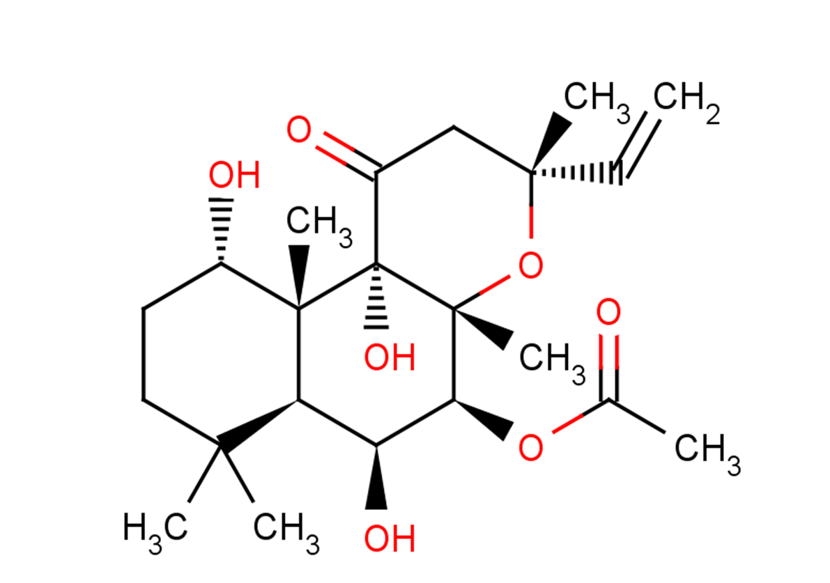
Forskolin 是从毛喉鞘蕊花中提取的一种天然产物,是腺苷酸环化酶激活剂,可以增加 cAMP 水平。它诱导多种细胞类型的分化并激活孕烷 X 受体和FXR,也可诱导细胞自噬。它对心脏产生正性肌力作用,具有血小板抗凝集和降压作用。
产品描述
Forskolin, a potent activator of the adenylate cyclase (EC50: 0.5 μM), can increase the cAMP level. It is extracted from the plant Coleus forskohlii.
体外活性
Forskolin (Fsk) treatment inhibited the proliferation of both Kit 225 and MT-2 cells in a dose-dependent manner with an IC50 equal to ~5 μM Forskolin. Fsk treatment (10–100 μM) increased cAMPi levels ~5- to 20-fold above basal levels, which reached maximum levels between 50–100 μM Fsk [2]. Forskolin binds to adenylyl cyclase in membranes from stably transfected Sf9 cells expressing type 1 adenylyl cyclase with an IC50 value of 41 nM and demonstrates an EC50 value of 0.5 μM in an activation assay assessing formation of cAMP from ATP [1]. "VC6T" [VPA, CHIR99021 (CHIR), 616452, Tranylcypromine] plus Forskolin (VC6TF) induced some GFP-positive clusters expressing E-cadherin [3].
体内活性
The Mrp4(-/-) mice exhibited no overt abnormalities in the development of the retinal vasculature, but retinal vascular development in the Mrp4(-/-) mice was suppressed in response to forskolin administration. The forskolin-treated Mrp4(-/-) mice showed an increased number of Ki67-positive and cleaved caspase 3-positive ECs, a significant decrease in the amount of pericyte coverage, and a reduced number of empty sleeves. In pups exposed to hyperoxia (75% oxygen) from P7 to P12, the Mrp4(-/-) mice showed a significant increase in the unvascularized retinal area [4]. Hepatic fibrosis induced by CCl4 was significantly reduced by forskolin, as indicated by decreased α-SMA expression and collagen deposition. Forskolin co-treatment significantly attenuated oxidative stress and inflammation, reduced TGF-β1 levels and down-regulated mRNA expression of Ptch-1, Smo and Gli-2 through cAMP-dependent PKA activation [5].
激酶实验
For Jak3 kinase assays, Fsk-treated MT-2 cells were lysed, clarified, and immunoprecipitated using Jak3 antibody as described above. Kinase reactions were carried out as described previously at 30 °C for 20 min. For PKA kinase assays, untreated MT-2 cells were lysed, and Jak3 was immunoprecipitated and bound to PAS beads as described previously. Immunoprecipitated Jak3 was washed with kinase buffer (50 mM Hepes-NaOH (pH 7.4), 10 mM MgCl2, 0.5 mM EGTA, 0.5 mM DTT, 20 μg/ml aprotinin, 10 μg/ml leupeptin, 1 μg/ml pepstatin A) and incubated with 200 μM ATP and purified protein kinase A catalytic subunit (PKAc) as indicated in the figure legends. Kinase reactions were carried out at 32 °C for 30 min followed by vigorous washing of the beads with cold kinase wash buffer as described previously. For [γ-32P]ATP radiolabeled kinase assays using recombinant Jak3, Hek293 cells were transfected with wild type (WT) Jak3 or kinase-dead Jak3 K855A using Lipofectamine 2000 according to the manufacturer's instructions. Cells were lysed and immunoprecipitated with Jak3 antibody. Jak3-bound PAS beads were washed three times in cold lysis buffer followed by kinase buffer. Kinase reactions were initiated by adding 10 μCi [γ-32P]ATP, 10 μm unlabeled ATP, and 1 μg of purified PKAc to Jak3-bound PAS bead reaction mixtures. Kinase reactions were performed at 32 °C for 30 min. Jak3-bound PAS beads were washed three times in radioimmunoassay buffer (10 mM Tris-HCl, pH 7.4, 75 mM NaCl, 20 mM EDTA, 10 mM EGTA, 20 mM Na4P2O7, 50 mM NaF, 20 mM 2-glycerolphosphate, 1 mM p-nitrophenyl phosphate, 0.1% Triton X-100) and one time in kinase wash buffer. The reactions were stopped by adding 2× SDS-PAGE sample buffer followed by SDS-PAGE. Coomassie stainable Jak3 bands were excised from the PVDF membrane and subjected to phosphoamino acid analysis [2].
细胞实验
Kit 225 or MT-2 cells were treated with 1, 5, 10, 20, 50, or 100 μM Forskolin for 20 min at 37 °C. Cells were lysed and clarified by centrifugation, and the concentration of cAMP was detected by direct cAMP ELISA. Optical density was measured at 405 nm, and the concentration of intracellular cAMP was calculated using a weighted four parameter logistic curve according to the manufactures instructions [2].
动物实验
Forskolin was dissolved in dimethyl sulfoxide (DMSO) and injected intraperitoneally into neonatal mice at postnatal days 4 (P4) and 5 (P5). Mice injected with DMSO served as the controls. The treated mice were euthanized at P6, and their retinas were isolated for whole-mount immunohistochemistry (IHC). We first tested the effect of different concentrations of forskolin on the survival rate and retinal vasculature and determined the optimal concentration, 1.0 μg/50 μL (0.3 mg/kg) at P4 and 1.5 μg/50 μL (0.5 mg/kg) at P5, used to compare the retinal vascular phenotypes between WT mice and Mrp4-deficient mice [4]. . After acclimatization for 2 weeks, animals were randomly divided into four groups of eight rats each and treated for six consecutive weeks as follows: The first group was treated with CCl4 (50% CCl4/corn oil; 0.5 mL·kg?1, i.p.) twice a week to induce liver fibrosis. The second group was given forskolin only at a dose of 10 mg·kg?1, i.p., dissolved in a DMSO/saline solution (1:49) five times a week. The third group was given both CCl4 and forskolin. The dose of forskolin used here was based on the results of our preliminary study. The fourth group served as the normal control, receiving vehicles only. At 24 h after the last injection, blood samples were collected from the retro‐orbital plexus after light anesthesia with sodium pentobarbital (50 mg·kg?1, i.p.). Serum was separated by centrifugation at 3000× g for 10 min and was used for the assessment of liver functions. Rats were killed by cervical dislocation, and livers were removed and weighed. A portion of liver tissue was washed and homogenized to obtain a 20% (w·v?1) homogenate, which was used for assessment of oxidative stress, inflammatory and fibrogenic markers. Another portion was placed in formalin for immunohistochemical and histopathological analyses. The remainder was stored at ?80°C, together with the 20% homogenate, until needed [5].
Cas No.
66575-29-9
分子式
C22H34O7
分子量
410.507
别名
Coleonol;毛喉素;Colforsin
储存和溶解度
Ethanol:15 mg/mL (36.5 mM)
H2O:Insoluble
DMSO:30 mg/mL (73 mM)
Powder: -20°C for 3 years
In solvent: -80°C for 2 years
 m.cnreagent.com