| CAS NO: | 3520-43-2 |
| 包装 | 价格(元) |
| 1mg | 电议 |
| 2mg | 电议 |
| 5mg | 电议 |
| 10mg | 电议 |
| 50mg | 电议 |
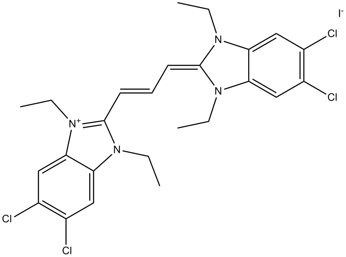
| Cas No. | 3520-43-2 |
| 别名 | CBIC2;JC1;JC 1 |
| 化学名 | 5,6-dichloro-2-[(E)-3-(5,6-dichloro-1,3-diethylbenzimidazol-3-ium-2-yl)prop-2-enylidene]-1,3-diethylbenzimidazole;iodide |
| Canonical SMILES | CCN1C2=CC(=C(C=C2[N+](=C1C=CC=C3N(C4=CC(=C(C=C4N3CC)Cl)Cl)CC)CC)Cl)Cl.[I-] |
| 分子式 | C25H27Cl4IN4 |
| 分子量 | 652.23 |
| 溶解度 | ≥ 32.6 mg/mL in DMSO with gentle warming |
| 储存条件 | Store at -20℃,protect from light |
| General tips | For obtaining a higher solubility , please warm the tube at 37 ℃ and shake it in the ultrasonic bath for a while. |
| Shipping Condition | Evaluation sample solution : ship with blue ice All other available size: ship with RT , or blue ice upon request |
| 产品描述 | JC-1 is a fluorescent lipophilic carbocyanine dye used to measure mitochondrial membrane potential. JC-1 (2.5 μM) exposed to murine L1210 lymphoblasts, can be detected the presence of both cytoplasmic JC-1 monomer and mitochondrial J-aggregates in these cells. JC-1 fluorescence is usually excited by the 488 nm laser wavelength common in flow cytometers[1]. Fluorescent labeling of mitochondria with either JC-1 (1 μg/mL, 15 min), reveals that are distributed irregularly, resulting in regions of high and low mitochondrial content within astrocytes[2]. JC-1 has been shown to interact with α-synuclein at the acidic C-terminal region with a Kd of 2.6 μM. JC-1 itself does not accelerate the protein aggregation of α-synuclein in the absence of iron, insted, it decelerates the aggregation process by extending the lag phase approx[3]. JC-1 is avidly accumulated in sensitive K562 cells where it displays both a green cytoplasmic and red mitochondrial fluorescence. JC-1 is poorly accumulated in resistant K562 cells, which displays only a slight green fluorescence[4]. References: |
 m.cnreagent.com