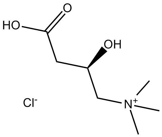
DL-Carnitine HCl 存在两种异构体,称为 D 和 L。
Kinase experiment: | Mitochondria (0.6 mg protein/mL) are incubated in 2.5 mM Hepes (pH7.4) containing 225 mM mannitol, 75 mM sucrose and 100 μM ethylene glycol tetraacetic acid (EGTA) with or without 5 mM L-carnitine at 25℃. To measure oxygen uptake, 10 min after inorganic phosphate (Pi) 4 mM are added, the mitochondria are treated with palmitoyl-CoA (50 μM) and then ADP is added (200 μM). Oligomycin (5 μM) and rotenone (10 μM) are added 3-4 min after the ADP treatment. HPG (0-10 mM), which can specifically inhibit carnitine palmitoyl transferase (CPT)-I activity in the mitochondria, is added in the Hepes medium before incubation of the mitochondria[1]. |
Animal experiment: | Rats: After 1 week of acclimatization, rats are randomly assigned to a hindlimb suspension group, hindlimb suspension with L-carnitine administration group, and a pair-fed group. The L-carnitine group are administered a 1250 mg L-carnitine/kg dissolved in distilled water orally using a sonde. The body weight is measured every morning at 09:00 and L-carnitine solution is ingested every morning at 10:00. The experiment is conducted for 14 days[3]. |
| 产品描述 | (±)-Carnitine chloride exists in two isomers, known as D and L. L-carnitine plays an essential role in the β-oxidation of fatty acids and also shows antioxidant, and anti-inflammatory activities.
The main role of L-carnitine is to shuttle long-chain fatty acids across the inner mitochondrial membrane. After L-carnitine and acyl-CoA become acyl-carnitine by activation of carnitine palmitoyl transferase (CPT)-I, the transported acyl-carnitine is changed into acyl-CoA by CPT-II in the mitochondria matrix. Palmitoyl-CoA-induced mitochondrial respiration is increased by L-carnitine treatment, and then is accelerated by the presence of ADP. This acceleration is induced by treatment with L-carnitine in a concentration-dependent manner, and is saturated at 5 mM L-carnitine[1]. Pretreatment with L-carnitine augments Nrf2 nuclear translocation, DNA binding activity and heme oxygenase-1 (HO-1) expression in H2O2-treated HL7702 cells. L-carnitine protects HL7702 cells against H2O2-induced cell damage through Akt-mediated activation of Nrf2 signaling pathway[2].
L-carnitine is found to down-regulate the ubiquitin proteasome pathway and increase IGF-1 concentrations in animal models. L-carnitine administration for 2 weeks of hindlimb suspension alleviates the decrease in weight and fiber size in the soleus muscle. In addition, L-carnitine suppresses atrogin-1 mRNA expression, which has been reported to play a pivotal role in muscle atrophy[3]. Simultaneous treatment with L-carnitine attenuates the renal fibrosis (which correlated with a reduction of plasma TGF-β1 levels) and the pro-oxidative and proinflammatory status reported in L-NAME groups, with a concomitant increase in the expression of PPAR-γ[4].
Reference:
[1]. Oyanagi E, et al. Protective action of L-carnitine on cardiac mitochondrial function and structure against fatty acidstress. Biochem Biophys Res Commun. 2011 Aug 19;412(1):61-7.
[2]. Li J, et al. l-carnitine protects human hepatocytes from oxidative stress-induced toxicity through Akt-mediated activation of Nrf2 signaling pathway. Can J Physiol Pharmacol. 2016 May;94(5):517-25.
[3]. Jang J, et al. l-Carnitine supplement reduces skeletal muscle atrophy induced by prolonged hindlimb suspension in rats. Appl Physiol Nutr Metab. 2016 Dec;41(12):1240-1247.
[4]. Zambrano S, et al. L-carnitine attenuates the development of kidney fibrosis in hypertensive rats by upregulating PPAR-γ. Am J Hypertens. 2014 Mar;27(3):460-70. |
 m.cnreagent.com