| 包装 | 价格(元) |
| 5mg | 电议 |
| 50mg | 电议 |
Cell lines | MEFs and HCT116 cells |
Preparation method | The solubility of this compound in DMSO is >10 mM. General tips for obtaining a higher concentration: Please warm the tube at 37 ℃ for 10 minutes and/or shake it in the ultrasonic bath for a while. Stock solution can be stored below - 20 ℃ for several months. |
Reacting condition | 0 ~ 2.5 μM; 8 hrs |
Applications | In both MEFs and HCT116 cells, 5-Iodotubercidin up-regulated p53 expression. Moreover, dosage experiments indicated that 5-Iodotubercidin was able to up-regulate p53 expression at the concentration as low as 0.25 μM. In HCT116 cells with ADK knocked out, the decrease of ADK levels did not significantly change the protein levels of p53, which indicated that 5-Iodotubercidin-induced p53 upregulation was not contributed to direct inhibition of ADK. |
Animal models | Nude mice bearing HCT116 cells |
Dosage form | 0.625 or 2.5 mg/kg; i.p. |
Applications | In nude mice bearing HCT116 cells, 5-Iodotubercidin at 2.5 mg/kg induced rapid tumor regression. However, 5-Iodotubercidin treatment also decreased the body weight of mice (a reduction of 6% at the end of treatment). Moreover, 5-Iodotubercidin showed inhibition on p53-/- HCT116-initiated tumors as well. At a lower dose of 0.625 mg/kg, 5-Iodotubercidin still showed an inhibition effect on tumor growth but p53-/- HCT116 exhibited resistance to 5-Iodotubercidin. |
Other notes | Please test the solubility of all compounds indoor, and the actual solubility may slightly differ with the theoretical value. This is caused by an experimental system error and it is normal. |
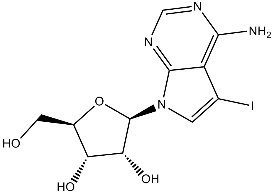
| 产品描述 | 5-Iodotubercidin (Itu) is a purine derivative and hence an inhibitor of adenosine kinase with an IC50 value of 26 nM [1]. Adenosine kinase is important in regulating the intracellular and extracellular concentrations of adenosine and hence diverse physiological actions of adenosine [2]. In various cells such as cancer cells, persisted AMPK activation could result in apoptosis [4]. In nude mice with colon carcinoma xenograft, Itu at a dose of 2.5 mg/kg resulted in rapid tumor regression compared with the control group. At the dose of 0.625 mg/kg, Itu still inhibited tumor growth, but p53-/- tumors were resistant to Itu at this lowered dose [1]. In male Wistar rat hepatocytes, incubation with Itu resulted in concentrations of AMP and ATP at 0.39 ± 0.06 and 1.51 ± 0.10 μmol/g cell wet mass, respectively; while control incubation at 0.27 ± 0.05 and 2.25 ± 0.33 μmol/g cell wet mass, respectively. Addition of 5-aminoimidazole-4-carboxamide ribonucleoside (AICAR) and Itu simultaneously resulted in almost the same effect of Itu alone. It was probable that Itu inhibited adenosine kinase and blocked the synthesis of 5-aminoimidazole-4-carboxamide ribonucleotide (ZMP) from AICAR. ZAM is a structural AMP analogue and hence mimics the effect of AMP on the AMP-activated protein kinase (AMPK) activation [3]. References: |
 m.cnreagent.com