| 包装 | 价格(元) |
| 50mg | 电议 |
| 100mg | 电议 |
| 250mg | 电议 |
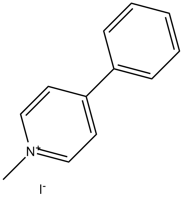
MPP+ 碘化物是神经毒素 MPTP 的有毒代谢物,通过选择性破坏黑质中的多巴胺能神经元,在动物模型中引起帕金森氏病症状。
Cell lines | Bv-2 cells |
Preparation Method | The cell experiment took Bv-2 cells as the object, set the MPP+ Iodidefinal concentration of 0.1, 0.2, 0.5 mmol as the interference concentration, and after 24 h of culture, Western blot detected the expression level of NLRP3 protein in cells, and selected the optimal concentration. |
Reaction Conditions | 0.1, 0.2, 0.5 mmol, 24h |
Applications | After 0.1/0.2/0.5 mmol MPP+ Iodide intervention cells for 24 h, MPP+ Iodideactivated cells expressed NLRP3 and MIF protein significantly higher than in the control group. 0.2 mmol MPP+ Iodideis the optimal concentration of NLRP3 inflammasomes that activate Bv-2. |
Animal models | Male Sprague–Dawley rats |
Preparation Method | Four days after siRNA infusion, rats were re-anesthetized for intranigral infusion of MPP+ Iodide(3 μg/μl) at a rate of 0.2 μl/min. After the surgery, rats recovered from anesthesia and were placed in home cages for the indicated times. |
Dosage form | 3 μg/μl;intranigral infusion |
Applications | The results shown intranigral infusion of MPP+ Iodideincreased HO-1 levels in a time-dependent manner; significant HO-1 elevation was observed 24 h to 7 d after MPP+ Iodideinfusion. |
| 文献引用 | |
| 产品描述 | MPP+ Iodide (1-methyl-4-phenylpyridinium iodide) is a toxic metabolite of the neurotoxin MPTP, and has successfully induced Parkinson-like syndromes in an in vitro model by selectively destroying dopaminergic neurons in substantia nigra.[1] In vitro efficacy test it shown that when SH-SY5Y cells were exposed to MPP+ Iodidein the range of 1–100 M for 3–24 h, MPP+ Iodide exhibited a dose-time dependent cytotoxicity.[1]In vitro experiment it indicated that SH-SY5Y cells were treated with 0.2, 0.4, 0.8, or 1.0 mM MPP + for 24 h, MPP+ Iodide could significantly reduce cell viability in a dose-dependent manner.[2]In vitro, treatment with 1-7.5 mM of MPP+ Iodide dose-dependently increased the neurodegeneration in the L1 larvae of BZ555 worms. The percentages of worms exhibiting neurodegeneration after treatment with 1 mM, 2.5 mM, 5 mM and 7.5 mM MPP+ Iodide were 24%, 27%, 67% and 87%, respectively.[3]Both TSM1 and primary neurons were treated with 0.1 to 2 mM of MPP+ Iodide induced neuronal cell death in a concentration dependent manner in vitro. TSM1 cells and primary neurons were treated with 400 μM MPP+ Iodide decreased by 60% and 80% the cell viability as compared to the control, respectively.[4]In vitro to test the role of MAC1 in MPTP/MPP+-induced neurotoxicity, neuron-glia cultures were treated with 0.125, 0.25, or 0.5 μM of MPP+ Iodidefound that MPP+-induced DAergic neurotoxicity in neuron-glia cultures was attenuated in the absence of MAC1.[5] In vivo study indicated that intranigral infusion of 3 μg/μl MPP+ Iodideinduced oxidative injury in nigrostriatal dopaminergic system of rat brain; and autophagy is pro-death in the MPP+-induced oxidative injury.[6] References: |
 m.cnreagent.com