| CAS NO: | 1403783-31-2 |
| 规格: | ≥98% |
| 包装 | 价格(元) |
| 5mg | 电议 |
| 25mg | 电议 |
| 50mg | 电议 |
| 100mg | 电议 |
| 250mg | 电议 |
| 500mg | 电议 |
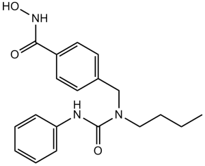
| Molecular Weight (MW) | 341.4 |
|---|---|
| Formula | C19 H23 N3 O3 |
| CAS No. | 1403783-31-2 |
| Storage | -20℃ for 3 years in powder form |
| -80℃ for 2 years in solvent | |
| Solubility (In vitro) | DMSO: 68 mg/mL (199.2 mM) |
| Water: <1 mg/mL | |
| Ethanol: 2 mg/mL (5.9 mM) | |
| Other info | Chemical Name: 4-((1-butyl-3-phenylureido)methyl)-N-hydroxybenzamide InChi Key: JZWXMCPARMXZQV-UHFFFAOYSA-N InChi Code: InChI=1S/C19H23N3O3/c1-2-3-13-22(19(24)20-17-7-5-4-6-8-17)14-15-9-11-16(12-10-15)18(23)21-25/h4-12,25H,2-3,13-14H2,1H3,(H,20,24)(H,21,23)SMILES Code: O=C(NO)C1=CC=C(CN(CCCC)C(NC2=CC=CC=C2)=O)C=C1 |
| Synonyms | Nexturastat A; AG-CR13901 |
| In Vitro | In vitro activity: In B16 murine melanoma cells, Nexturastat A leads to a dose-dependent increase of acetyl α-tubulin levels without a concomitant elevation of histone H3 acetylation. Moreover, Nexturastat A potently inhibits the growth of B16 melanoma cells with GI50 of 14.3 μM. Kinase Assay: HDAC inhibition assays are performed by Reaction Biology Corp. using isolated human, recombinant full2length HDAC1 and -6 from a baculovirus expression system in Sf9 cells. An acetylated fluorogenic peptide,RHKKAc, derived from residues 379-382 of p53 is used as substrate. The reaction buffer is made up of 50 mM Tris-HCl pH 8.0, 127 mM NaCl, 2.7 mM KCl, 1 mM MgCl2, 1 mg/mL BSA, and a final concentration of 1% DMSO. Compounds are delivered in DMSO and delivered to enzyme mixture with preincubation of 5-10 min followed by substrate addition and incubation for 2 h at 30°C. Trichostatin A and developer are added to quench the reaction and generate fluorescence, respectively. Dose-response curves are generated starting at 30 μM compound with three-fold serial dilutions to generate a 10-dose plot. IC50 values are then generated from the resulting plots, and the values expressed are the average of duplicate trials ± standard deviation. Cell Assay: B16 murine melanoma cells are plated at 5000/well in 96 well flat bottom plates. The following day, media is changed to that containing various concentrations of HDACi or matched DMSO vehicle concentrations diluted in complete medium done in triplicate. Cells are incubated for 48 hours at 37°C and 5% CO2. Density of viable, metabolically active cells is quantified using a standard MTS assay as per manufacturer’s instructions. Briefly, 20μL of reagent are added per well and incubated at 37°C for 3 hours. Absorbances at 490nM are measured spectrophotometrically with background subtraction at 690 nM. All values are then normalized and expressed as a percentage of medium control (100%). |
|---|---|
| In Vivo | NA |
| Animal model | NA |
| Formulation & Dosage | NA |
| References | J Med Chem. 2012 Nov 26;55(22):9891-9. |
 m.cnreagent.com