| CAS NO: | 927961-18-0 |
| 规格: | ≥98% |
| 包装 | 价格(元) |
| 5mg | 电议 |
| 10mg | 电议 |
| 25mg | 电议 |
| 50mg | 电议 |
| 100mg | 电议 |
| 250mg | 电议 |
| 500mg | 电议 |
| Molecular Weight (MW) | 434.0162 |
|---|---|
| Formula | C19H15ClN2O4S2 |
| CAS No. | 927961-18-0 |
| Storage | -20℃ for 3 years in powder form |
| -80℃ for 2 years in solvent | |
| Solubility (In vitro) | DMSO:>100 mg/mL |
| Water: <1 mg/mL | |
| Ethanol: <1 mg/mL | |
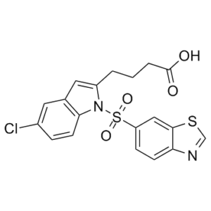
| SMILES Code | O=C(O)CCCC(N1S(=O)(C2=CC=C3N=CSC3=C2)=O)=CC4=C1C=CC(Cl)=C4 |
| Synonyms | IVA-337; IVA337; IVA 337; Lanifibranor |
| In Vitro | In vitro activity: In vitro Caco2 permeability assessment: Compounds were tested at 10 μM in a 96-well permeable plate seeded with Caco-2 cells. The medium was the same in apical and basolateral sides: Hanks’ Balanced Salt Solution (HBSS) + 5 mM Hepes + bovine serum albumin 1%, pH 7.4. The assay was performed with a robotic platform (Caliper-Perkin Elmer system). After incubation for 2 hours, the concentrations of tested compound was measured in both sides by LC/MS /MS (API4000 Qtrap, AB Sciex). Permeability in both directions (apical to basolateral and basolateral to apical) was assessed to determine the efflux ratio. Kinase Assay: PPAR Transactivation assays: These cell-based assays were carried out using Cos-7 cells transfected with a chimeric human or murine PPARα-Gal4 receptor expression plasmid (or PPARδ-Gal4, or PPARγ-Gal4) and a 5Gal4 pGL3 TK Luc reporter plasmid. Transfections were performed by a chemical agent (Jet PEI). Transfected cells were distributed in 384-wells plates and were allowed to recover for 24 h. The culture medium was then removed and replaced by fresh medium containing the compounds to be tested (variable concentration in 0.5% DMSO). After an overnight incubation, luciferase expression was measured by adding SteadyGlo according to the manufacturer’s instructions (Promega). Fenofibric acid at 10-5 M (PPARα), GW501516 at 10-8 M (PPARδ), and rosiglitazone at 10-6 M (PPARγ) were used as references. Results were expressed as fold induction compared to basal level or as percentage activity compared to references taken as 100%. Calculation and plate validation from run to run were done using the software Assay Explorer (MDL). Serial dilutions of compounds (final concentration ranging from 30 μM to0.001 μM) were tested in triplicate on an automated screening core-system from Beckman or Caliper. The EC50 calculation was done using Assay Explorer (MDL) and was determined simultaneously on human and mouse PPARα/δ/γ. Cell Assay: Beta-oxidation assays: The functional PPARα and PPARδ activity was determined in two cell lines, HuH7 (human liver hepatoma cells, JCRB 0403) and C2C12 (mouse muscle cells differentiated into myotubes, ATCC CRL-1772) as measurement of oleate beta-oxidation. Cells were seeded into Petri dishes with a central well and incubated in DMEM medium at 37°C. Compounds were added in DMSO (0.1% final concentration) at, at least, 3 different concentrations for 48 hours. Two hours before the end of the incubation period, albumin-bound 14C-oleate was added in the cell medium culture. The reaction was stopped by addition of a 40% perchloric acid solution to remove excess 14C-oleate in the medium. Given off CO2 was trapped by KOH in the central well after sealing of the dishes, for 90 minutes, at room temperature and then counted. Compounds were tested in triplicate for each concentration. Data are expressed as % of variation vs GW501516 used as reference compound at 0.1 μM. |
|---|---|
| In Vivo | The animals were housed in groups of 3-10 in polypropylene cages (floor area = 1032 cm2) under standard conditions: room temperature (22 ± 2°C), hygrometry (55 ± 10%), light/dark cycle (12h/12h), air replacement (15 - 20 volumes/hour), water and food (SDS, RM1) ad libitum. Mice were allowed to habituate for at least 5 days prior to experimentation. Mice were numbered by marking their tail using indelible markers The study was conducted under EU animal welfare regulation for animal use in experimentation (European Directive 2010/63/EEC). This experimental protocol was submitted for approval by the Inventiva Ethical Committee “Comité de reflexion Ethique en Expérimentation Animale (CR2EA) (registered by the “Ministère de l’Enseignement Supérieur et de la Recherche” under No. 104). All the procedures described below were reviewed and approved by the Inventiva ethics committee. Inventiva is a company AAALAC fully accredited. In vivo animal model of diabetic dyslipidemia: db/db mice In vivo animal model of fibrosis: CCl4 C57Bl6/J mice In vivo safety model: in Sprague-Dawley rats. Sprague Dawley rats (6 weeks of age) were fed with a standard pellet diet or a diet supplemented with compounds at a low or a high dose. Tested compounds were 5 (100 and 1000 mg/kg), 1a (3 and 30 mg/kg), 3a (10 and 100 mg/kg) and 3b (1 and 10 mg/kg). Plasma volume was measured by Evans blue dye dilution. Briefly, conscious animals were briefly restrained in a commercial restrainer for tail vein injection of 1 mg of Evans blue dye (200 μL in saline). After 10 min, 0.5 mL of blood was withdrawn from orbital sinus under light anesthesia. A small amount was withdrawn into two hematocrit capillaries and centrifuged for 5 min at 12,000 g to measure hematocrit. The remainder of the sample was centrifuged for 10 min at 3,500 g and dye concentration was determined in the plasma. The absorbance measured at 620 nm was compared with a standard curve generated by using a pool of blood from 3 untreated rats. |
| Animal model | Mouse; Homozygous C57BL/Ks-db male mice (db/db mice); C57Bl6/J mice; Sprague Dawley rats |
| Formulation & Dosage | Please refer to the above 'in vivo' section' for more information about the different formulations and dosages associated with the different in vivo models |
| References | J Med Chem. 2018 Mar 22;61(6):2246-2265. |
 m.cnreagent.com